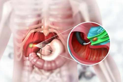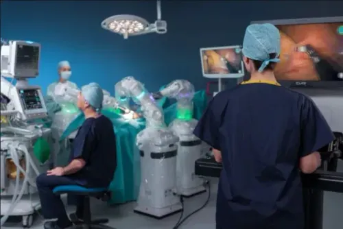
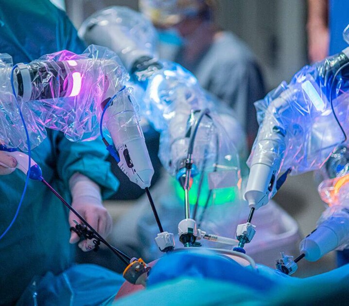
Esophageal surgery involves more than placing instruments through small incisions: the team must confirm the diagnosis, select the correct operation, mobilise the esophagus, remove disease when necessary and rebuild the passage for food. You will learn what happens before, during and after surgery, which tests and risks matter, and how to assess a robotic surgeon and hospital serving patients near Nelamangala.
Key takeaways
- The diagnosis, tumour stage and anatomy determine the operation—not the robot.
- Expect imaging, endoscopy, biopsy review and fitness tests before surgery.
- Ask how robotic, laparoscopic, open and endoscopic options compare for your condition.
- Confirm the team’s experience, hospital support and recovery plan near Nelamangala.
Which esophageal operation fits your condition?
Diagnosis determines the operation, not the robot. A robotic surgeon for esophageal conditions considers whether you have cancer, achalasia, severe confirmed reflux, a benign stricture or another problem, then matches treatment to tumour location, stage, anatomy, previous operations, lung function and overall fitness.
| Condition or approach | What the operation involves | When it applies |
|---|---|---|
| Esophagectomy | Removing the diseased esophagus, reshaping the stomach into a conduit and joining it to the remaining esophagus | Selected cancers or severe damage |
| Transhiatal approach | Removing the esophagus through abdominal and neck access without opening the chest | Tumour location and anatomy permit this route |
| Transthoracic approach | Adding chest access for esophageal mobilisation and lymph-node removal | Tumours requiring chest dissection |
| Ivor Lewis or McKeown | Ivor Lewis joins the stomach in the chest; McKeown joins it in the neck | Reconstruction after cancer resection |
| Heller myotomy | Cutting tight lower-esophageal muscle, usually with an anti-reflux procedure | Achalasia |
| Anti-reflux surgery | Reconstructing the valve at the stomach entrance | Objectively confirmed reflux after suitable testing |
Cancer care may combine chemotherapy, radiation, chemoradiation before surgery, immunotherapy and surgery. Stage, tumour biology and fitness determine the sequence; surgery is not automatically first.
An esophageal surgery consultation should also discuss nutrition and recovery. Ask which operation fits your anatomy and lung function, and whether prior surgery changes the plan. A robotic approach cannot make an uncertain reflux diagnosis appropriate.
What tests happen before an esophageal surgery consultation?
A normal endoscopy does not answer every question. An esophageal surgery consultation needs tests that identify the disease, map its extent, and show whether you can safely undergo treatment.
- Upper endoscopy with biopsy: The camera examines the lining, and tissue confirms or excludes suspected cancer. An endoscopy report alone cannot show tumour spread or determine the complete operation.
- Contrast CT of the chest and abdomen: CT maps the esophagus, nearby organs, enlarged lymph nodes, and possible distant disease.
- PET-CT: Use this for cancer staging when the oncology team considers it indicated; it can reveal metabolically active disease outside the primary site.
- Endoscopic ultrasound: This assesses tumour depth through the esophageal wall and nearby lymph nodes, sometimes allowing targeted needle sampling.
- Barium swallow or timed barium esophagram: Swallowed contrast shows narrowing, anatomy, and how quickly food or liquid leaves the esophagus.
- High-resolution esophageal manometry: This confirms and classifies achalasia after structural obstruction has been excluded.
- Reflux monitoring: pH or impedance testing objectively confirms reflux before anti-reflux surgery, especially when symptoms could reflect achalasia or functional disorders.
- Pulmonary function tests, blood tests, ECG, or echocardiography: Use these when lung or heart fitness needs assessment.
A multidisciplinary review brings together gastrointestinal, thoracic, oncology, radiology, pathology, anaesthesia, and nutrition expertise. If you are comparing robotic esophageal surgery nelamangala options, ask who reviews all results together, not just the endoscopy.
How the robotic operation is performed step by step
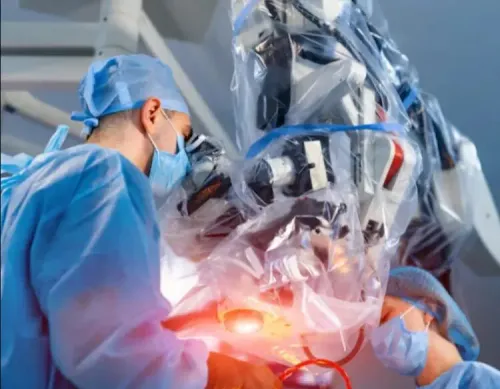
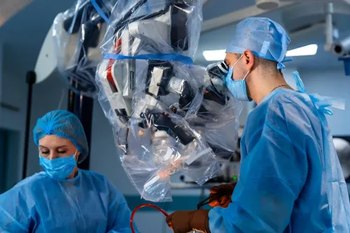
Admission includes fasting, medication review, consent, and nutritional planning before general anaesthesia. After you are asleep, the team positions you to provide access to the abdomen and chest, or the neck when required, while protecting pressure points and monitoring breathing.
1. The surgeon places small ports in the abdomen and chest, or abdominal and neck ports for a three-stage operation. A camera and robotic instruments enter through these ports.
2. Seated at the console, the surgeon controls the instruments to free the esophagus from surrounding tissue. For cancer, the team removes nearby lymph nodes for pathological staging.
3. The surgeon removes the diseased esophageal segment and reshapes the stomach into a narrow conduit, which is the usual replacement. Selected patients need colon interposition instead.
4. The team joins the conduit to the remaining esophagus and places a feeding tube to support nutrition while swallowing recovers. The operation can be changed to an open procedure if bleeding, dense scar tissue, anatomy, or safety requires it.
| Reconstruction | Join location | Usual pattern |
|---|---|---|
| Ivor Lewis | Inside the chest | Abdominal and chest stages |
| McKeown | In the neck | Abdominal, chest, and neck stages |
Ivor Lewis and McKeown esophagectomy therefore differ mainly by anastomosis location and surgical stages. A search for robotic surgeon esophageal surgery near Nelamangala or robotic esophageal surgery Nelamangala does not prove suitability: the surgeon chooses the approach, cut line, and reconstruction. The robot neither diagnoses nor operates independently.
How robotic, laparoscopic, open and endoscopic treatment differ
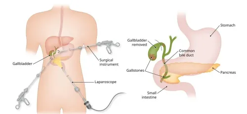
Small ports do not mean a small operation. Robotic and laparoscopic esophageal surgery enter through several abdominal or chest ports; open surgery uses a larger chest or abdominal incision; endoscopic treatment reaches the esophagus through the mouth without abdominal or chest access.
| Option | Access and strengths | Trade-offs |
|---|---|---|
| Robotic | Surgeon-controlled instruments through ports, with magnified three-dimensional vision and articulated wrists | Limited tactile feedback; requires experienced port placement and dissection |
| Laparoscopic | Two-dimensional camera view through small ports and conventional instruments | Less instrument articulation, though the incisions are smaller than open surgery |
| Open | Direct access through a larger incision | More wound pain and recovery burden, but useful for difficult anatomy, bleeding or extensive disease |
| Endoscopic | Flexible instruments pass through the mouth | Suits selected inner-wall problems, not removal of an esophageal cancer segment |
A robotic surgeon for esophageal conditions still makes every decision: the robot does not diagnose, choose the cut line or operate independently. Smaller incisions do not remove esophagectomy risks such as pneumonia, bleeding, anastomotic leak, narrowing, recurrent laryngeal-nerve injury, chylothorax or cardiac complications.
Conversion to open surgery is prudent when bleeding cannot be controlled safely, scar tissue obscures anatomy, the tumour cannot be removed with clear margins, or exposure is inadequate. It is a safety decision, not a failure of robotic esophageal surgery nelamangala.
Treatment names also matter:
- Heller myotomy, usually paired with anti-reflux surgery, divides tight muscle for achalasia.
- Peroral endoscopic myotomy divides that muscle through an endoscope.
- Dilation stretches a narrowing; it does not remove cancer.
- Cancer resection removes the diseased esophagus and requires reconstruction.
What recovery involves and how to choose the right surgical team near Nelamangala
After surgery, you may spend time in intensive care or a monitored ward. Nurses track oxygen, heart rate, drains and urine; the team controls pain, teaches deep-breathing exercises, and gets you walking early to reduce pneumonia and blood clots. Imaging, drain output and clinical changes help detect an anastomotic leak.
A feeding tube supplies nutrition while the join heals. A swallowing assessment precedes the staged progression below:
| Stage | Focus | What you receive |
|---|---|---|
| Tube feeding | Protect healing join | Nutrition through the feeding tube |
| Clear liquids | Test swallowing | Small, supervised sips |
| Soft foods | Build tolerance | Frequent, moist, easy-to-chew meals |
| Wider diet | Restore intake | Smaller meals, gradual food expansion |
Reflux, weight loss, dumping symptoms and narrowing at the join need follow-up; narrowing can require endoscopic dilation. Hospitalisation varies by operation and recovery. Do not drive while taking opioid pain medicine or unable to brake safely; return to work depends on strength and job demands.
- Anastomotic leak, bleeding, infection, pneumonia, respiratory failure and blood clots.
- Recurrent laryngeal-nerve injury, chyle leak and prolonged feeding-tube use.
After discharge, seek urgent advice for fever, worsening breathlessness, chest pain, persistent vomiting, inability to swallow liquids, increasing abdominal or neck pain, a racing pulse, wound discharge or sudden leg swelling.
- Where will surgery occur, how long is travel, and who reviews you afterward?
- Is intensive care available, with emergency thoracic and gastrointestinal specialists?
- What upper-GI training and experience does the robotic surgeon for esophageal conditions have with your proposed operation?
Robotic Cancer Surgery should explain the complete referral pathway, not only the platform.
Related service
ROBOT ASSISTED ESOPHAGEAL SURGERY ROBOT ASSISTED ESOPHAGEAL SURGERY Robot-assisted esophageal surgery provides precision and minimally invasive techniques for treating esophageal conditions such as cancer, achalasia,... View service → |
Frequently asked questions
Which esophageal operation fits my condition?
The choice depends on the diagnosis, tumour location and stage, anatomy, previous operations, lung function and overall fitness. Cancer, achalasia, severe confirmed reflux and benign strictures require different treatments.
What tests happen before an esophageal surgery consultation?
Your evaluation can include endoscopy, biopsy review, CT or PET-CT imaging, contrast studies, oesophageal manometry and tests of lung and general fitness. The exact set depends on your diagnosis.
How is robotic esophageal surgery performed?
The surgeon places instruments through small abdominal or chest incisions, controls them from a console and removes or repairs the affected area. The operation may include lymph-node removal and reconstruction when cancer treatment requires it.
How do robotic, laparoscopic, open and endoscopic treatment differ?
Endoscopic treatment works through the oesophagus, laparoscopic and robotic surgery use small incisions, and open surgery uses a larger incision. The safest approach depends on the condition, anatomy and complexity of the procedure.
How do I choose a surgical team near Nelamangala?
Ask about the surgeon’s experience with your specific operation, the hospital’s intensive-care and endoscopy support, expected recovery, complication management and who will coordinate follow-up.
Keywords